The Regenerex System has been shown in many clinical trials to successfully close up to 95% of non-responding chronic wounds within 90 days. The wounds clinically tested had already been subject to current protocols of treatment and failed to heal. Competitive clinical trials indicated there isn’t another System that has the ability to close chronic wounds at these levels and speed. Not only does our System close chronic wounds relatively quickly but it does so at a very reasonable cost.
Our QBx™ technology is the most efficacious, and clinically proven, chronic wound care product line in the world. Chronic wounds are those that fail to progress through a normal, orderly, and timely sequence of repair. They are not only characterized by delayed healing for weeks, months, or even years, but also by a resistance to treatment with conventional dressings and therapies. They impart a particularly devastating financial and quality-of-life burden on individuals suffering from the wounds and are frustrating for the caregivers and clinicians who attempt to manage, but fail to heal these wounds.
Non-healing chronic wounds are thought to be a consequence of factors that affect both the production of new tissue and the elevated destruction of existing tissue. Biochemically, these wounds appear to be stuck in a catabolic, inflammatory phase that is hostile to local growth factors and the activity of fibroblasts and keratinocytes. Increases in matrix metalloproteinases (MMPs) MMP-2 and MMP-9 are of significance in non-healing chronic wounds.
MMPs are a group of zinc-containing proteolytic enzymes that play an important role in the remodeling of the extracellular matrix of wounds. An overproduction of MMPs may result in degradation of the extracellular matrix and inactivation of vital growth factors. A precisely orchestrated balance of MMP production and their natural inhibitors (TIMPs) is needed.
Additionally, it is theorized that wounds stuck in the inflammatory phase persistently overproduce free radicals or reactive oxygen species (ROS). At low concentration and early in the inflammatory phase of wound healing, ROS such as hydrogen peroxide (H2O2), have a positive effect on healing through stimulation of fibroblast proliferation. However, persistent overproduction of ROS is thought to be detrimental to healing. A new treatment strategy has emerged focused upon manipulating the expression of genes which control the endogenous production of MMPs and TIMPs within the local wound environment. Contrary to modalities designed to sequester MMPs and/or act as a competitive substrate for protease activity, this technology strategy relies on delivery of metal ions into the wound to help regulate gene expression for the production of MMPs and TIMPs, thus bringing them into balance. These metal ions are delivered via a polyethylene glycol based, QBx™ ointment, which also contains citric acid to help normalize wound pH and reduce ROS activity. The QBx™ ointment is delivered via a tube or an acetylated regenerated cellulose carrier which allows for the passage of wound drainage and is non-fibre shedding. The entire composition is marketed as our primary wound dressing called Accelerex™.
Approximately 80% of chronic wounds display elevated levels of MMPs. Traditionally, however, these wounds have been managed with simple gauze and gauze-like dressings to cover and protect the wound. Other modern wound dressings such as hydrocolloids and collagens absorb wound fluids, but these dressings do not impact the cellular environment. This void in the treatment regimen offers a unique market advantage. QBx™ contributes to creating a suitable environment to allow wounds to close. These formularies are based on Proteases Down Regulating Technology and provide the System for a comprehensive suite of wound care products focused on the treatment of chronic wounds.
Wound Types
Acute Wounds
Acute wounds can occur from traumatic abrasions, burns, lacerations, and superificial skin and soft tissue injuries.
Diabetic Foot Ulcers
Diabetic Foot Ulcers are a common chronic wound for people with diabetes.
Venous Ulcers
Venous Ulcers are the most common and costly chronic wound ulcer in the US and affect women three times more often than men.
Case Studies
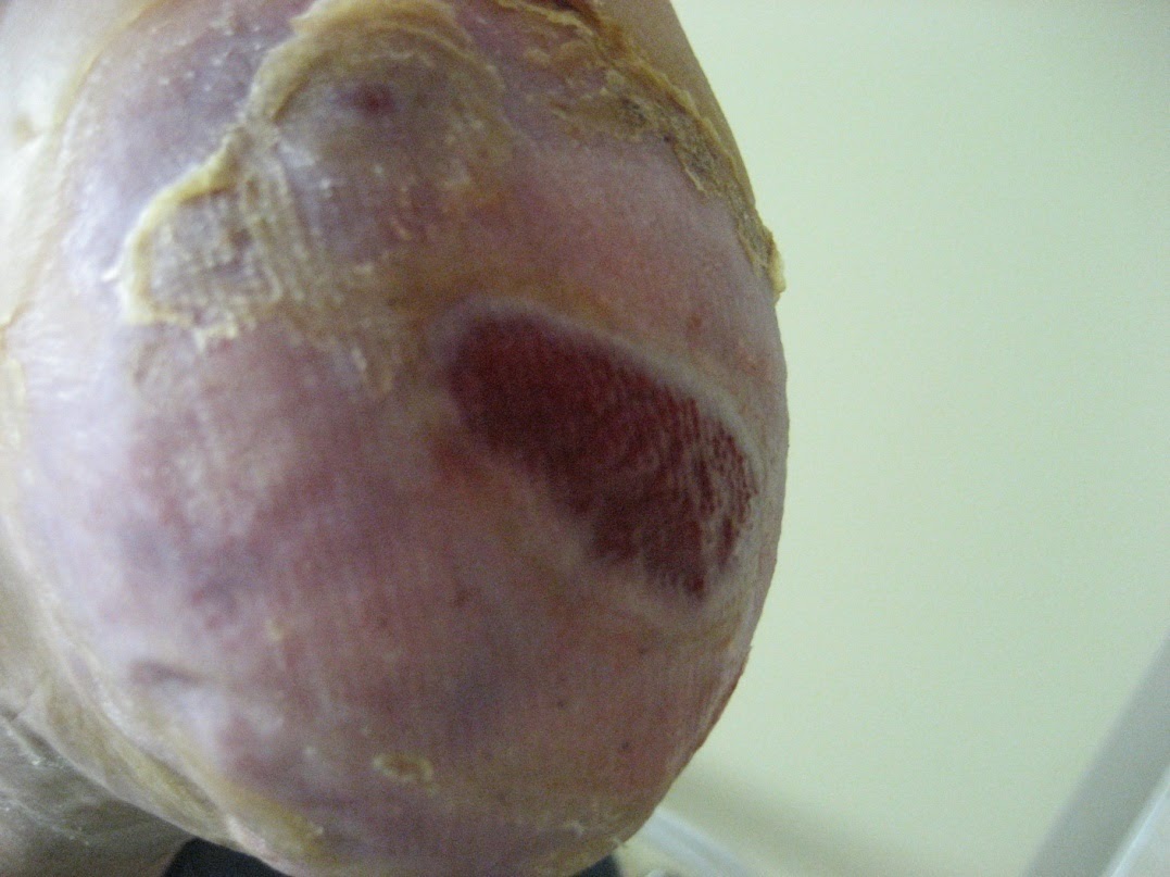
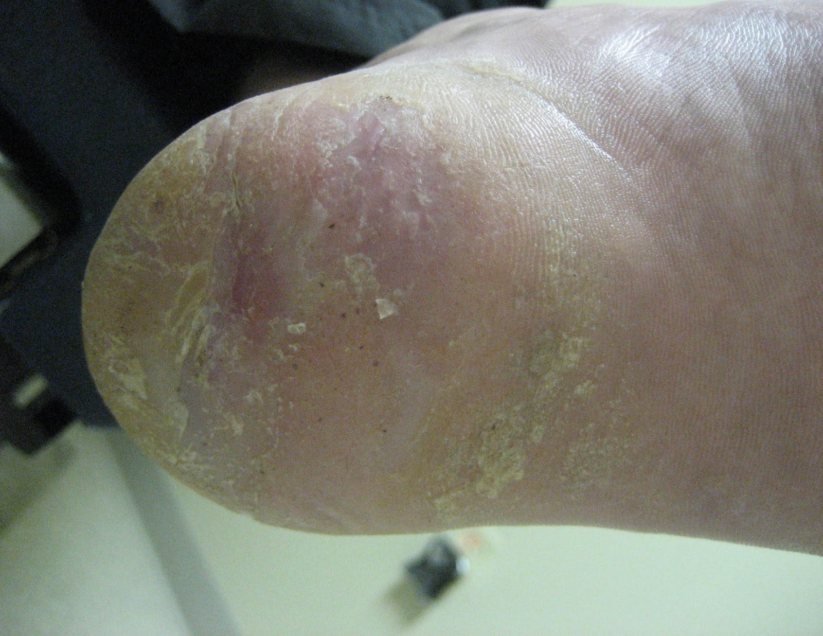
Diabetic ulcer
In existence for 3 months prior to treatment
Week 0
Week 7
2nd/3rd degree electrical burns
Earlier failed treatments: Elasto-Gel, SSD (Flammazine)
Week 0
Week 7
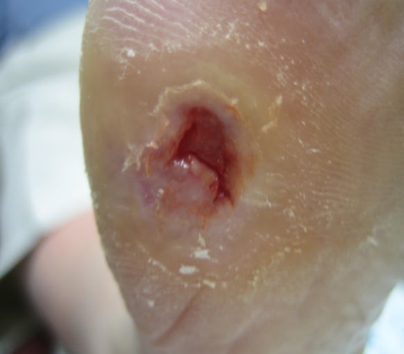
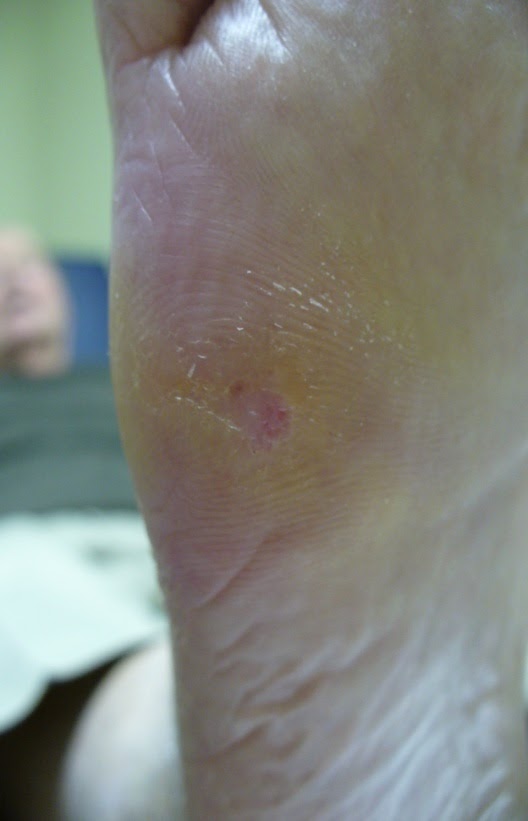
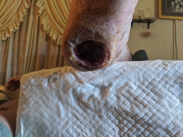
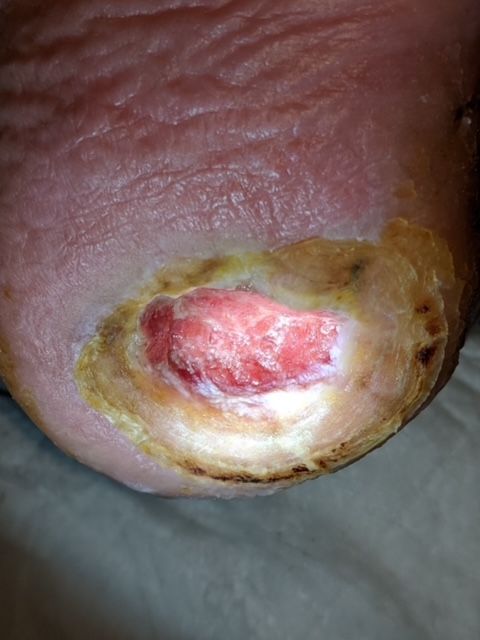
Diabetic ulcer
60 year old diabetic male
Week 0
Week 13
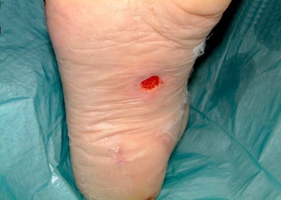
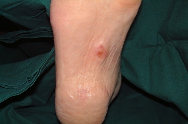
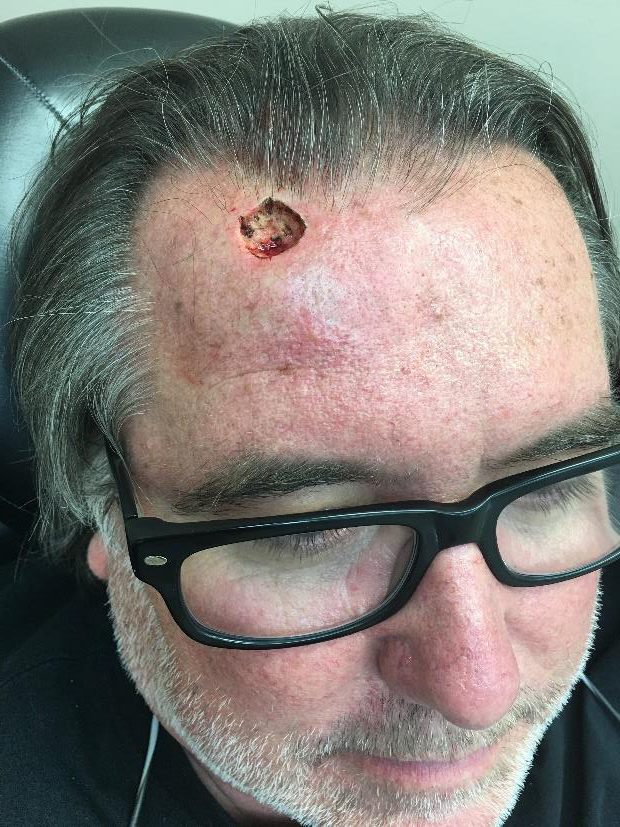
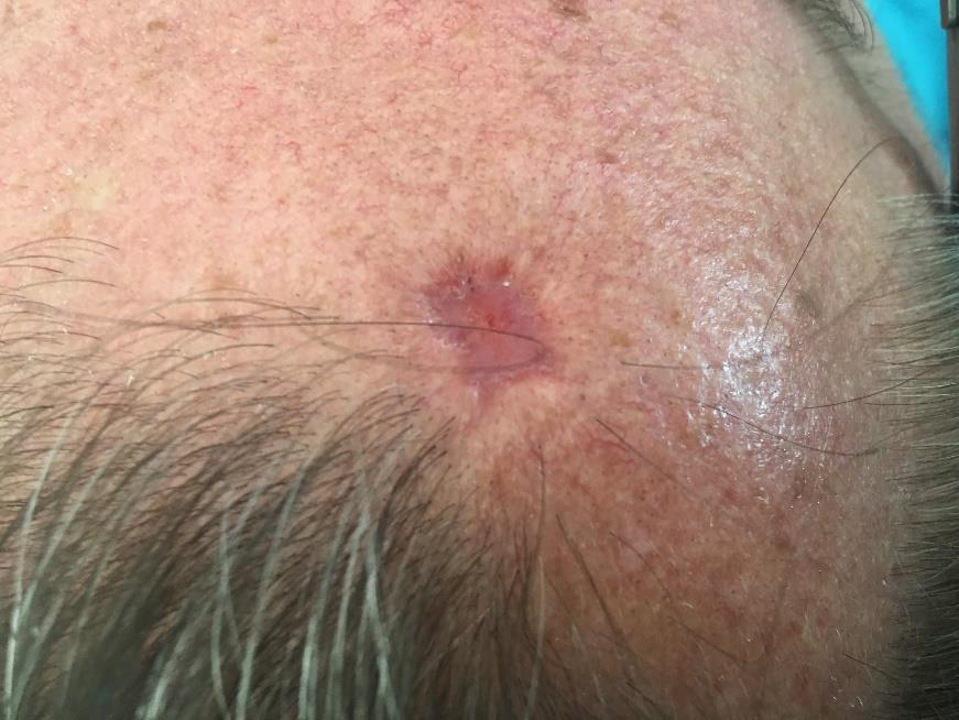
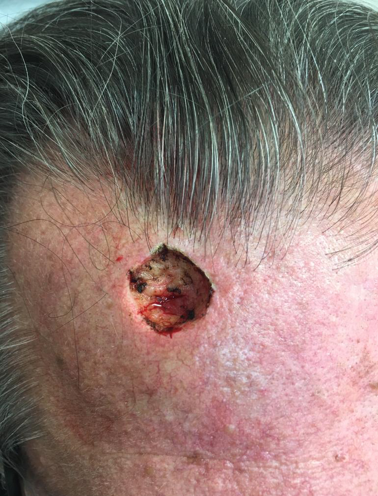
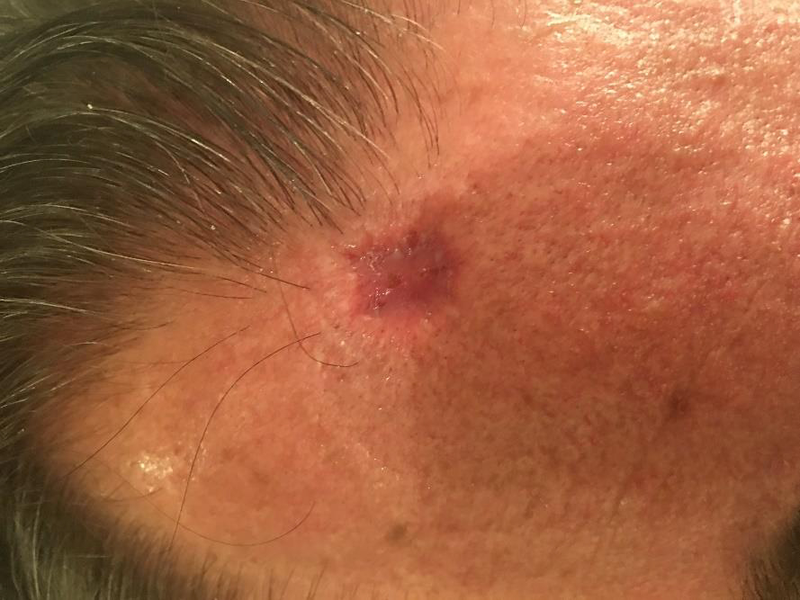
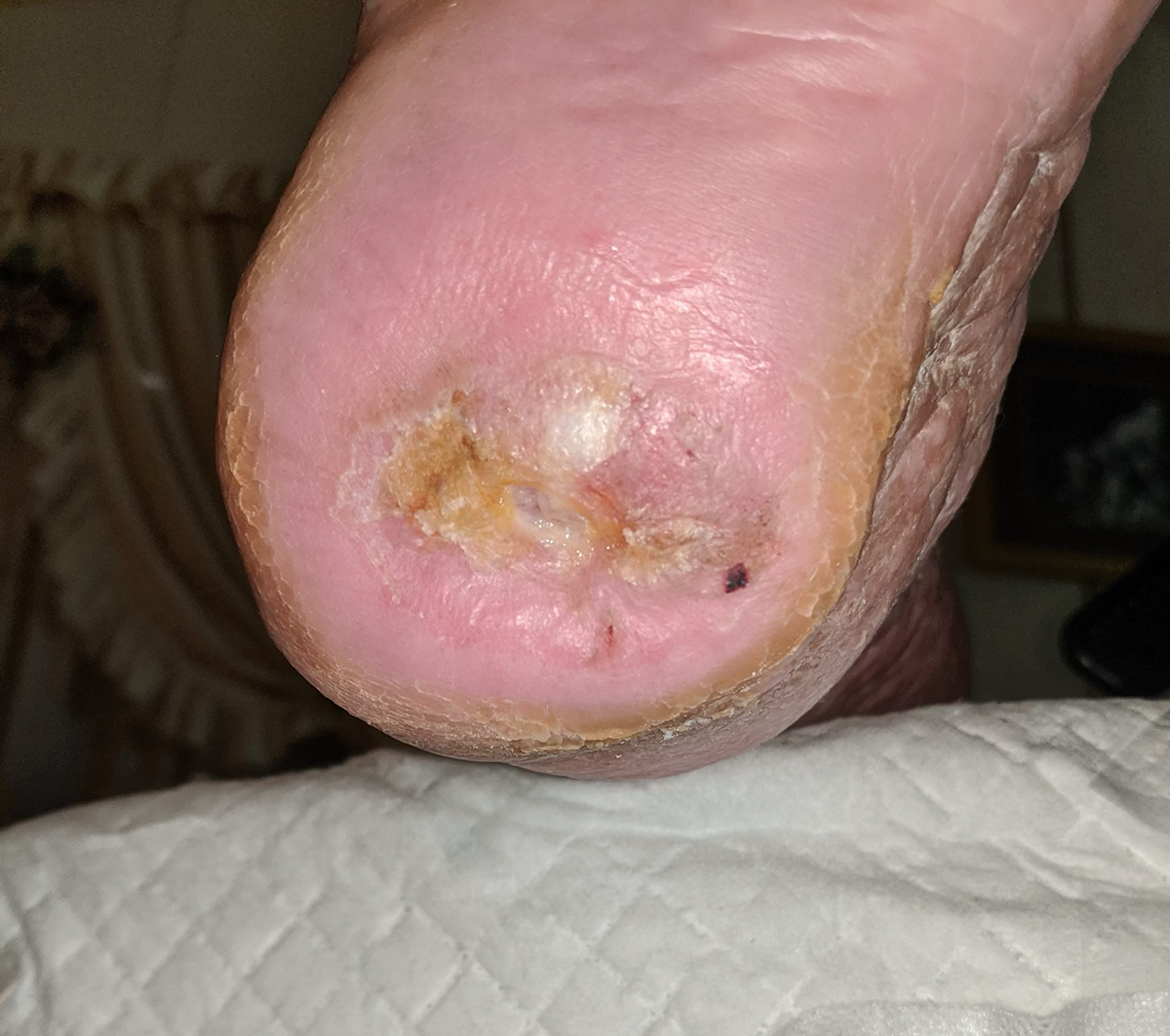
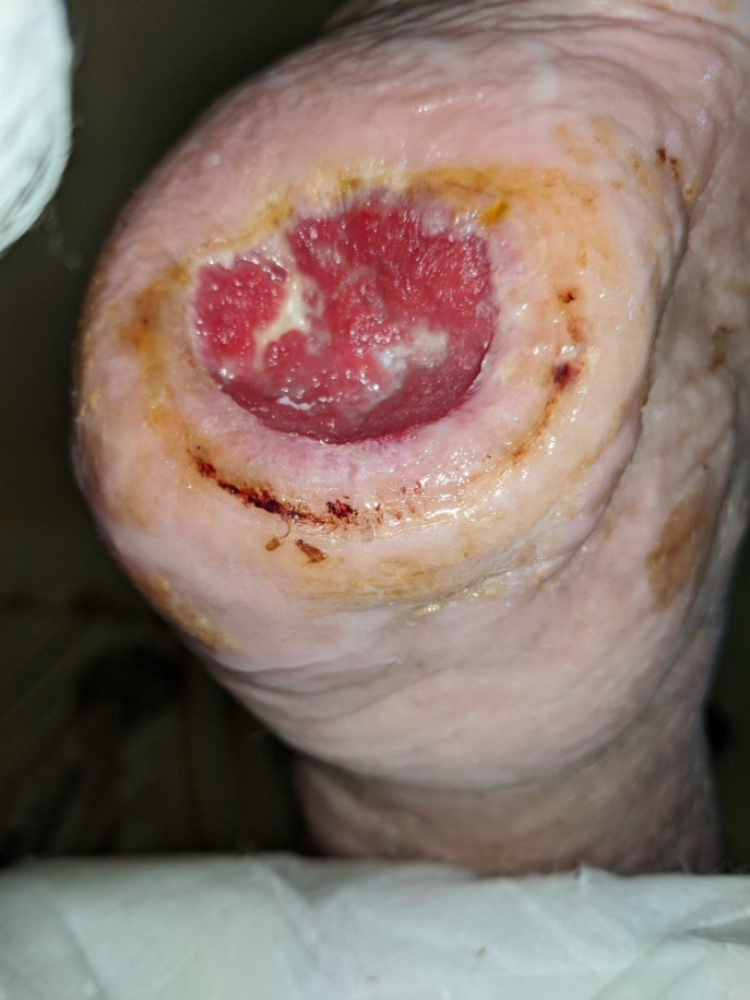
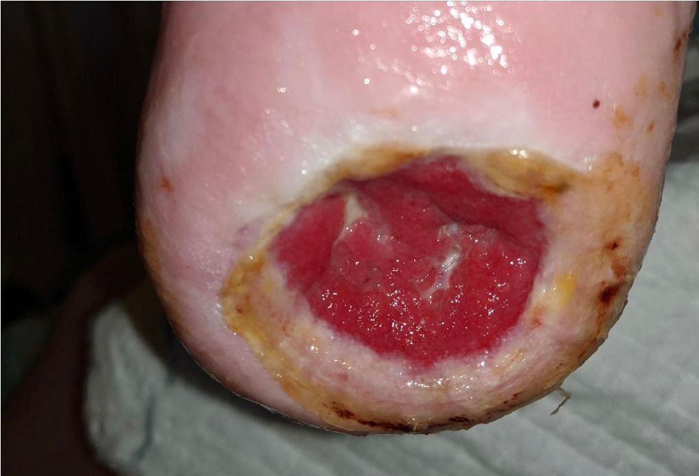
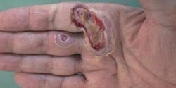
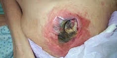
Cancer removal surgery
The wound length, width and depth measurement was 2.58,2.68, & .71 centimeters.
Day 0
Day 17
Day 0
Day 17
Diabetic ulcer
Week 0
Week 13
Diabetic ulcer
Week 0
Week 6
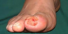
Diabetic ulcer
2 cm x 2.8 cm wide and 1 cm deep before treatment
Week 0
Week 2
Week 5
Week 2
Week 2
Week 5
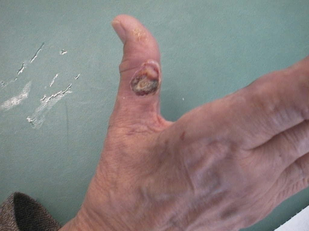
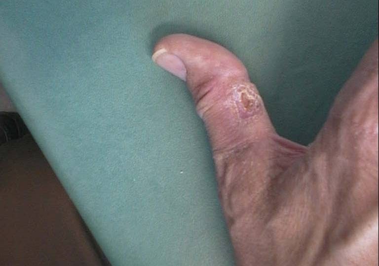
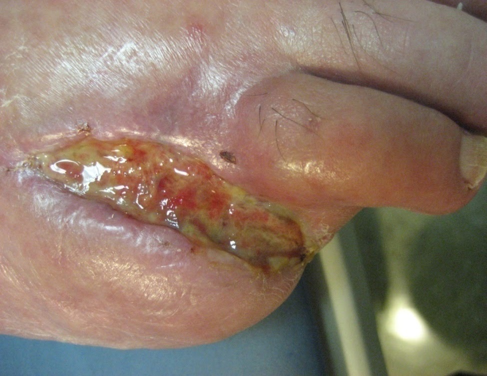
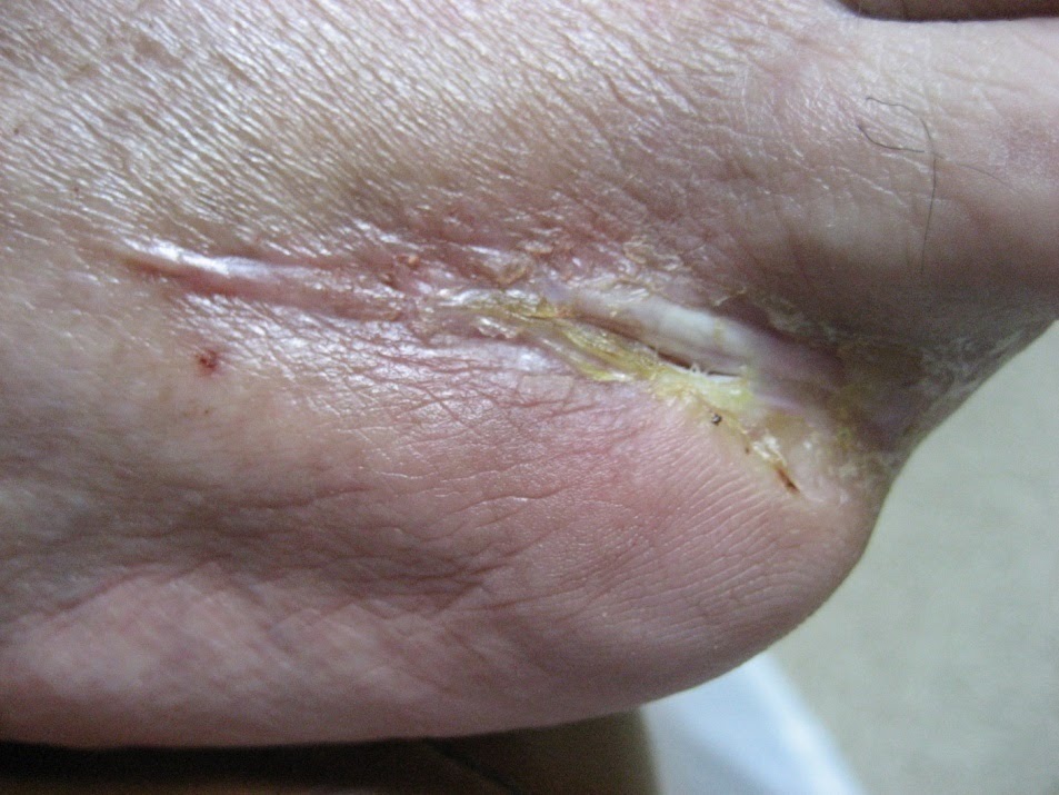
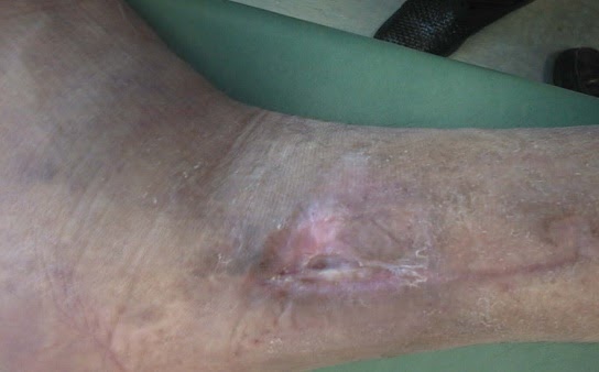
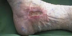
Post-traumatic ulcer
In existence for over a year; earlier failed treatment: Kaltostat
Week 0
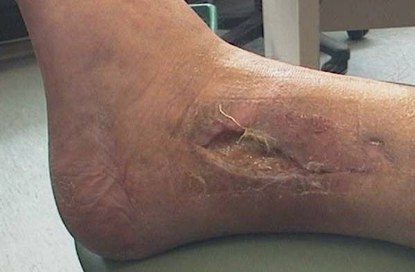
Week 7
Wound Information
Acute Wounds
Acute wounds can occur from traumatic abrasions, burns, lacerations, and superificial skin and soft tissue injuries. (Source 1 – Kane 2003) Acute wounds tend to heal spontaneously without complications through the four phases of wound healing which are hemostasis, inflammation, proliferation, and maturation.
Chronic Wounds
A chronic wound is a wound that fails to heal within a six to twelve week period of best clinical practice. (Source 2) They are a challenge to both healthcare professionals and patients because they last a long time causing a loss of body image and make the person more susceptible to infection. Several factors can impact the normal healing process such as infection, necrotic tissue, impaired tissue perfusion and steroids can all contribute to non-healing wounds. (Source 3)
Chronic wounds can occur anywhere on the body including the lower extremities. They are commonly referred to as diabetic foot ulcers , venous stasis ulcers and pressure ulcers. Chronic wounds fail to progress through an organized, orderly and timely sequence of wound repair. The patients involved may have other pre-existing medical conditions such as diabetes mellitus that could contribute to the non-healing, chronic wound.. It is important to determine the underlying etiology of the chronic wound and identify any underlying factors that interfere with the healing process such as malnutrition, poor glucose control, excessive pressure to an area, anemia and persistent edema. Elderly patients who are experiencing a chronic wound may have multiple medical conditions that necessitate a non-surgical approach to wound healing. Acceleration of wound closure is important for individuals with chronic wounds because they are at risk for infection, and these wounds can interfere with activities of daily living.
The common types of chronic wounds are as follows:
Diabetic Foot Ulcers (DFU)
Diabetic foot ulcers are a significant problem on a global basis. Approximately 15% of people with diabetes will develop a foot ulcer and it is estimated that more than half of these will experience a second ulcer. Diabetic foot ulcers occur most commonly due to neuropathy (loss of sensation in the lower extremity) and peripheral vascular disease. Neuropathy can occur partially as a complication of prolonged glucose elevation (Steed 2007) Failure of diabetic foot ulcers to heal may result in amputation.
The main reasons for diabetic foot ulcers failing to heal are two-fold. One primary cause is that the person with the foot ulcer continues to walk on the ulcer causing irritation and skin breakdown. They may also have some type of foreign body enter the skin and be unaware of this happening. For the healing process to take place, these wounds usually have weight redistributed away from the wound in what is known as offloading. Specialty shoes and devices are available to assist with this process.
The second main reason that these ulcers fail to heal is related to proper diabetes control. Uncontrolled blood sugar can prevent the body from healing itself by preventing certain healing process from occurring. Management includes prevention of weight bearing (off loading) , debridement of devitalized tissue, prevention of infection and local wound management.
Venous Ulcers
Venous Ulcers are the most common and costly chronic wound ulcer seen in the United States and affect women three times more often than men. The prevalence of lower limb ulcers ranges from 0.12% to 0.32% of the general population. (Burrows 2006) Patients experiencing venous ulcers suffer from the inability to heal and the high rate of reoccurrence at 72%. (Sibbald 2006) Venous ulcers can cause pain, limit activities of daily living and negatively impact quality of life.
Venous ulcers can be caused by damaged or leaky venous valves, or a faulty calf muscle pump action which causes to sustained high venous pressure known as venous hypertension. These ulcers are usually located around the medial malleolus and are accompanied by edema or swelling of the affected leg, large amounts of drainage, and often have a scaly type of skin condition known as dermatosis. It is important to note that not all leg ulcers are venous in origin particularly those ulcers located above or below the gaiter and ankle region.
People with venous insufficiency sometimes report having had a blood clot in the affected limb. Due to the interruption of blood flow back to the heart, pooling of blood is seen in the limb that sometimes results in swelling. Eventually, protein from blood vessels can leak into the tissue and cause an ulcer to form. A cornerstone of treatment for venous ulcers beyond local ulcer management is the use of sustained compression in order to decrease the edema by wrapping the legs to promote blood flow back to the heart.
Pressure Ulcers
Pressure ulcers are also known as decubitus ulcers or bed sores have a psycologic, physiologic and economic impact on the individual. Pressure ulcers can result from immobility and can occur in persons that are bed ridden, who have had a spinal cord injury, or are in a wheelchair. Prevalence and incidence across all care settings varies ranging from 0.4%-38% in general acute care, 2.2% to 23.9% in long term care, and 0% to 17% in home care. (Source: Weir 2007) The tissue injury that occurs sometimes is a result of sustained pressure to a bony prominence area such as the hip or heel. The sustained pressure prevents adequate blood flow and causes the tissue to die.
The wounds often fail to heal for a variety of reasons. One main reason is that pressure is not relieved from the wound. Special mattresses, bed and seating cushions are designed to help to relieve pressure to allow better blood flow to the injured area. Other reasons for these wounds to fail to heal often have to do with other underlying medical conditions that affect individuals, such as poor nutrition and incontinence, shear and friction. Pressure ulcer management includes pressure relief/pressure reduction, proper positioning, prevention of infection and local wound management.
References
1 Krasner D,Rodeheaver, G and Sibbald RG. Chronic Wound Care:A clinical source book for healthcare professionals;4th edition.HMP Communications 2007;page 14.
2 Kunimoto B, Cooling M, Gulliver W, Houghton P, Orsted H, Sibbald RG, Best Practices for the prevention and treatement of venous leg ulcers.Ostomy Wound Management 2001;47 (2):34-46,48-50.
3 McCance KL and Heather SE.Pathophysiology:The biologic basis for disease in adults and children. 3rd ed. Mosby,St. Louis, 1998:232-234.
4 Krasner D,Rodeheaver, G and Sibbald RG. Chronic Wound Care:A clinical source book for healthcare professionals;4th edition.HMP Communications 2007; chapter 53;page 537.
5 Krasner D,Rodeheaver, G and Sibbald RG. Chronic Wound Care:A clinical source book for healthcare professionals;4th edition.HMP Communications 2007;page chapter 44;page 429
6 Krasner D,Rodeheaver, G and Sibbald RG. Chronic Wound Care:A clinical source book for healthcare professionals;4th edition.HMP Communications 2007; chapter 44;page 429.
7 Krasner D,Rodeheaver, G and Sibbald RG. Chronic Wound Care:A clinical source book for healthcare professionals;4th edition.HMP Communications 2007; chapter 58; page 575.